Lined by Non-keratinized stratified squamous epithelium
Anterior side
Layngeal mucosa:
Lined by Psuedostratified squamous epithelium
Lamina propria with serous and mucinous glands
Central core of elastic cartilage
Larynx
Fig.
False vocal cord:
Lined by Pseudostratified ciliated columnar epithelium with goblet cells.
In lamina propria, are numerous and mixed seromucous glands.
Ventricle:
Lined by Pseudostratified ciliated columnar epithelium.
Lamina propria blends with the perichondrium of the hyaline thyroid cartilage.
True vocal cord:
Lined by Stratified squamous epithelium
Thin, dense lamina propria devoid of glands, lymphatic tissue, or blood vessels.
Trachea
Fig.
Consists of 4 layers:
Mucosa
Lined by Pseudostratified ciliated columnar epitheliium.
Lamina propria contains fine connective tissue fibres, diffuse lymphatic tissue, and occasionaly solitary lymphatic nodules.
Located deeper in the lamina propria is the longitudinal elastic membrane that separates the lamina propria from submucosa.
Submucosa
Submucosa contains the loose connective tissue as that of mucosa.
Hyaline cartilage and trachealis muscle
Hyaline cartilage is surrounded by perichondrium.
Adventitia
Numerous nerves, blood vessels and adipose tissue are located in the adventitia.
Bronchus
Fig.
Lining: Pseudostratified ciliated columnar epithelium with goblet cells
Submucosa contains both serous and mucous acini
Smooth mucles present between mucosa and cartilage
Cartilage present in irregular patches.
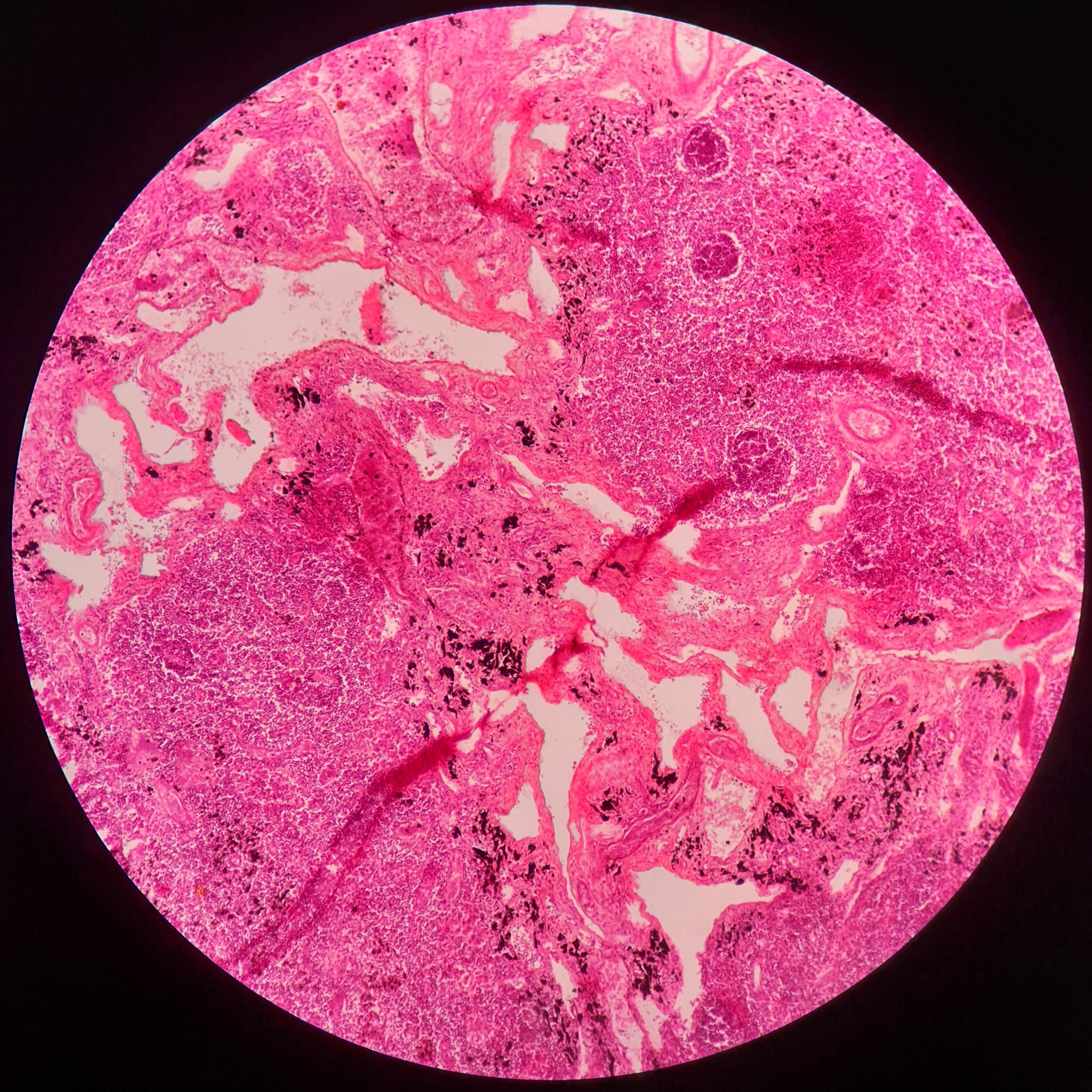
Lungs
Fig.
Fig.
Presence of alveolar ducts.
Alveolar sac lined by simple squamous epithelium.
Presence of intrapulmonary bronchus with varying amount of islands cartilage and smooth muscles.
Respiratory bronchioles with simple cuboidal epithelial lining lacking cilia and goblet cells.
Cardiovascular
Elastic artery
Fig.
Fig.
Tunica intima consisting of endothelium, subendothelial connective tissue, and internal elastic lamina.
The first layer of elastic fibers is called the internal elastic lamina. The internal elastic lamina is not distinct from the elastic fibers of media.
Well developed subendothelial layer in tunica intima.
Thick tunica media with many elastic fibers and some smooth muscle fibers.
Tunica adventitia containing collagen fibers with several elastic fibers.
Vasa vasorum in the tunica adventitia.
Muscular artery
Fig.
In muscular arteries, the tunica intima is made up of endothelium and internal elastic lamina, which is thrown into wavy folds due to contraction of smooth muscle in the media.
Tunica media is composed mainly of smooth muscle fibers arranged circularly.
Tunica adventitia contains collagen fibers and few elastic fibers.
Inferior Venacava
Fig.
Venacava
Fig.
The vein has a thinner wall and a larger lumen than the artery.
The tunica intima, media, and adventitia can be made out, but they are not sharply demarcated.
The media is thin and contains a much larger quantity of collagen fibers than arteries. The amount of elastic tissue or of muscle is much less.
The adventita is relatively thick and contains considerable amount of elastic and muscle fibers.
Lymphatic System
Thymus
Fig.
The thymus is made up of lymphoid tissue arranged in the form of distinct lobules. The presence of this lobulation enables easy distinction of the thymus from all other lymphoid organs
The lobules are partially separated from each other by connective tissue septae In each lobule an outer darkly stained cortex (in which lymphocytes are densely packed); and an inner lightly stained medulla (in which the cells are diffuse) are present
Whereas the cortex is confined to one lobule, the medulla is continuous from one lobule to another
The medulla contains pink staining rounded masses called the corpuscles of Hassall.
Spleen
Fig.
The spleen is characterized by a thick capsule with trabeculae extending from it into the organ (not shown in photomicrograph)
The substance of the organ is divisible into the red pulp in which there are diffusely distributed lymphocytes and numerous sinusoids; and the white pulp in which dense aggregations of lymphocytes are present. The latter are in the form of cords surrounding arterioles
When cut transversely the cords resemble the lymphatic nodules of lymph nodes, and like them they have germinal centers surrounded by rings of densely packed lymphocytes.
However, the nodules of the spleen are easily distinguished from those of lymph nodes because of the presence of an arteriole in each nodule
Lymph nodes
Fig.
Fig.
A thin capsule surrounds the lymph node and sends in trabeculae
Just beneath the capsule a clear space is seen. This is the subcapsular sinus
A lymph node has an outer cortex and an inner medulla
The cortex is packed with lymphocytes. A number of rounded lymphatic follicles (or nodules) are present. Each nodule has a pale staining germinal center surrounded by a zone of densely packed lymphocytes
Tonsil
Fig.
Palatine tonsil is an aggregation of lymphoid tissue that is readily recognized by the fact that it is covered by a stratified squamous epithelium
At places the epithelium dips into the tonsil in the form of deep crypts
Deep to the epithelium there is diffuse lymphoid tissue in which typical lymphatic nodules can be seen.
Digestive system
Tongue
Fig.
The tongue is covered on both surfaces by stratified squamous epithelium (nonkeratinized)
The ventral surface of the tongue is smooth, but on the dorsum the surface shows numerous projections or papillae
Each papilla has a core of connective tissue covered by epithelium. Some papillae are pointed (filiform), while others are broad at the top (fungiform).
A third type of papilla is circumvallate, the top of this papilla is broad and lies at the same level as the surrounding mucosa
The main mass of the tongue is formed by skeletal muscle seen below the lamina propria.
Muscle fibers run in various directions so that some are cut longitudinally and some transversely
Numerous serous and mucous glands are present amongst the muscle fibers.
Circumvallate papillae
Fig.
Circumvallate papillae are characterized by their dome-shaped structure lined by stratified squamous epithelium
Numerous oval-shaped lightly stained taste buds can be seen on the lateral wall of the papillae
The underlying connective tissue contains serous glands of von Ebner
Skeletal muscle can be seen extending into the papillae.
Oesophagus
Fig.
Four layers of gastrointestinal tract (GIT): Mucosa, submucosa, muscularis externa, adventitia seen
Lining epithelium is stratified squamous nonkeratinized epithelium
Submucosa is studded with mucus-secreting esophageal glands.
Stomach
Fig.
Presence of four layers—mucosa, submucosa, muscularis externa, and serosa
Deep gastric pits occupying two-thirds of the depth of the mucosa
Presence of pyloric glands (mucous glands) in the mucosa that are simple/branched tubular glands which are coiled.
Duodenum
Fig.
Wall made up of four layers
Mucosa—presence of numerous broad villi, lined by simple columnar epithelium with microvilli and goblet cells
Presence of submucous Brunner’s gland.
Jejunum
Fig.
Wall made up of four layers
Mucosa—presence of numerous tall slender villi, lined by simple columnar epithelium with microvilli and goblet cells
Absence of submucous Brunner’s gland.
Ileum
Fig.
Wall made up of four layers
Mucosa—presence of short, small fewer villi, lined by simple columnar epithelium with microvilli and numerous goblet cells
Absence of submucous Brunner’s gland.
Presence of Peyer's patch
Appendix
Fig.
Wall made up of four layers
Mucosa—absence of villi, lined by simple columnar epithelium with numerous goblet cells
Submucosa packed with numerous lymphatic follicles, extending into lamina propria
Absence of taenia coli, appendices epiploicae.
Colon
Fig.
Wall made up of four layers
Mucosa—absence of villi, lined by simple columnar epithelium with numerous goblet cells
Lymphatic follicles dispersed in the lamina propria
Presence of taenia coli, appendices epiploicae.
Gall bladder
Fig.
The mucous membrane is lined by tall columnar cells with striated border
The mucosa is highly folded and some of the folds might look-like villi
Crypts may be found in lamina propria Submucosa is absent
The muscle coat is poorly developed with numerous connective tissue fibers interspersed among muscle. This is called as fibromuscular coat
Liver
Fig.
The panoramic view of liver shows many hexagonal areas called hepatic lobules.
The lobules are partially separated by connective tissue
Each lobule has a small round space in the center. This is the central vein
A number of broad irregular cords of cells seem to pass from this vein to the periphery of the lobule. These cords are made up of polygonal liver cells—hepatocytes
Along the periphery of the lobules, there are angular intervals filled by connective tissue
Each such area contains a branch of the portal vein, a branch of the hepatic artery, and an interlobular bile duct
These three constitute a portal triad.
Pancreases
Fig.
Made up of serous acini
The cells forming the acini of the pancreas are highly basophilic (bluish staining). The lumen of the acinus is very small
Some acini may show pale staining (centroacinar cell) in the center
Among the acini, some ducts are seen
The ducts have a distinct lumen, lined by cuboidal epithelium
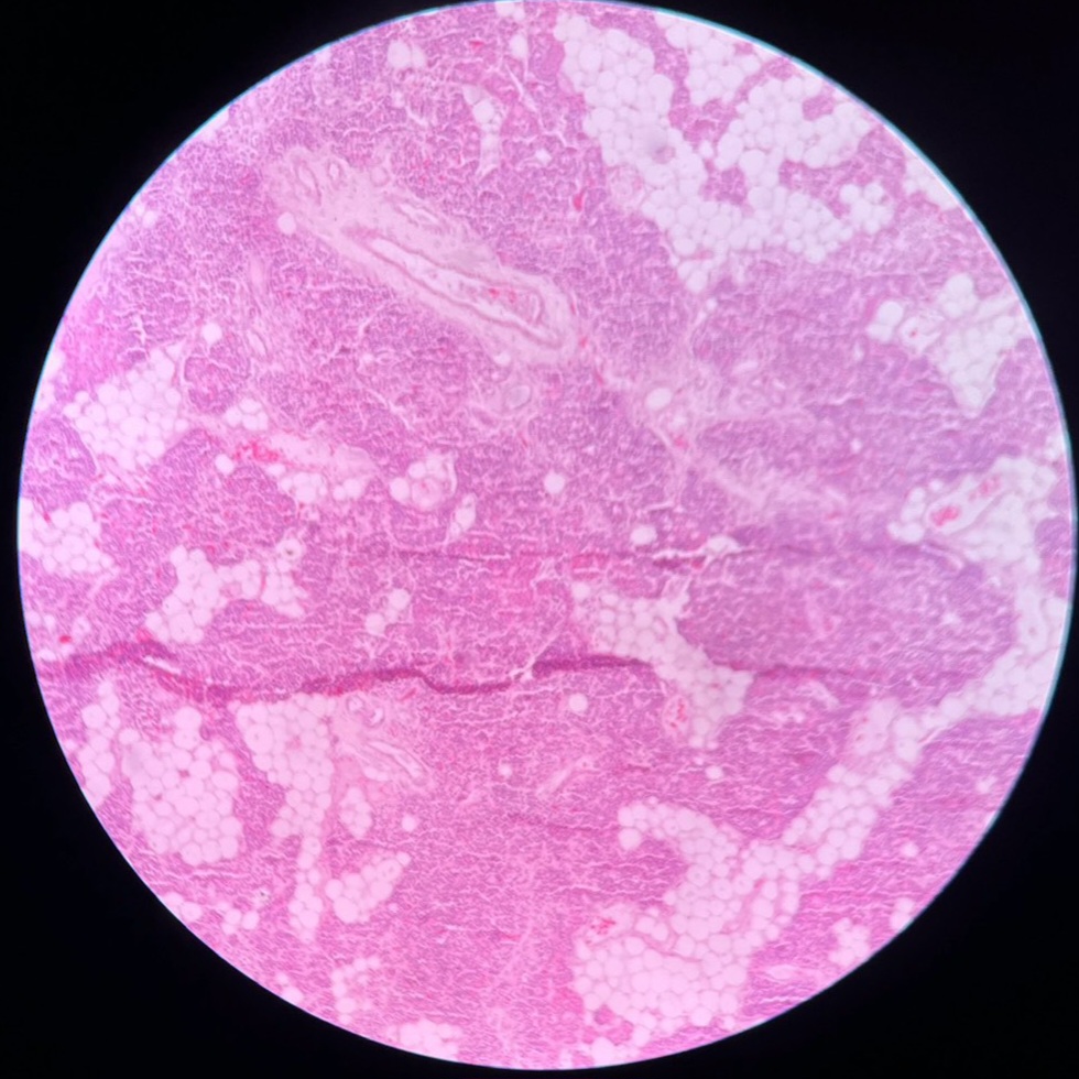
Parotid gland
Fig.
The parotid gland is a serous salivary gland.
The characteristic features are:
Only serous acini are present which contain basophilic zymogen granules and are darkly stained Intercalated and striated (intralobular) ducts are seen
Interlobular duct can be seen It also contains adipocytes.
Submandibular gland
Fig.
The submandibular gland is a mixed salivary gland, predominantly serous with a few mucous acini
Serous cells are frequently located at the periphery of mucous acini in the form of a crescent and called as demilunes
Striated ducts are more prominent than those in parotid gland.
Sublingual gland
Fig.
The sublingual gland is predominantly a mucous gland but few serous acini may also be seen
Serous demilunes may be present.
Endocrine system
Pituitary gland
Fig.
The hypophysis cerebri consists of three main parts:
Pars anterior is cellular
Pars intermedia is variable in structure
Pars posterior consists of fibers, and is lightly stained.
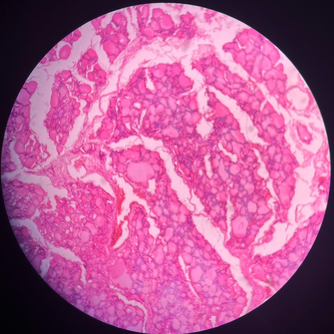
Thyroid gland
Fig.
The thyroid gland is made up of follicles lined by cuboidal epitheliumIn photomicrograph, in low magnification, it can be seen that follicles vary in shape and size
Each follicle is filled with a homogenous pink colloid proteinaceous material composed primarily of thyroglobulin that has been produced by the follicular epithelial cells
Parafollicular cells are present in relation to the follicles and also as groups in the connective tissue
In the intervals between the follicles, there is some connective tissue and blood vessels between follicles.
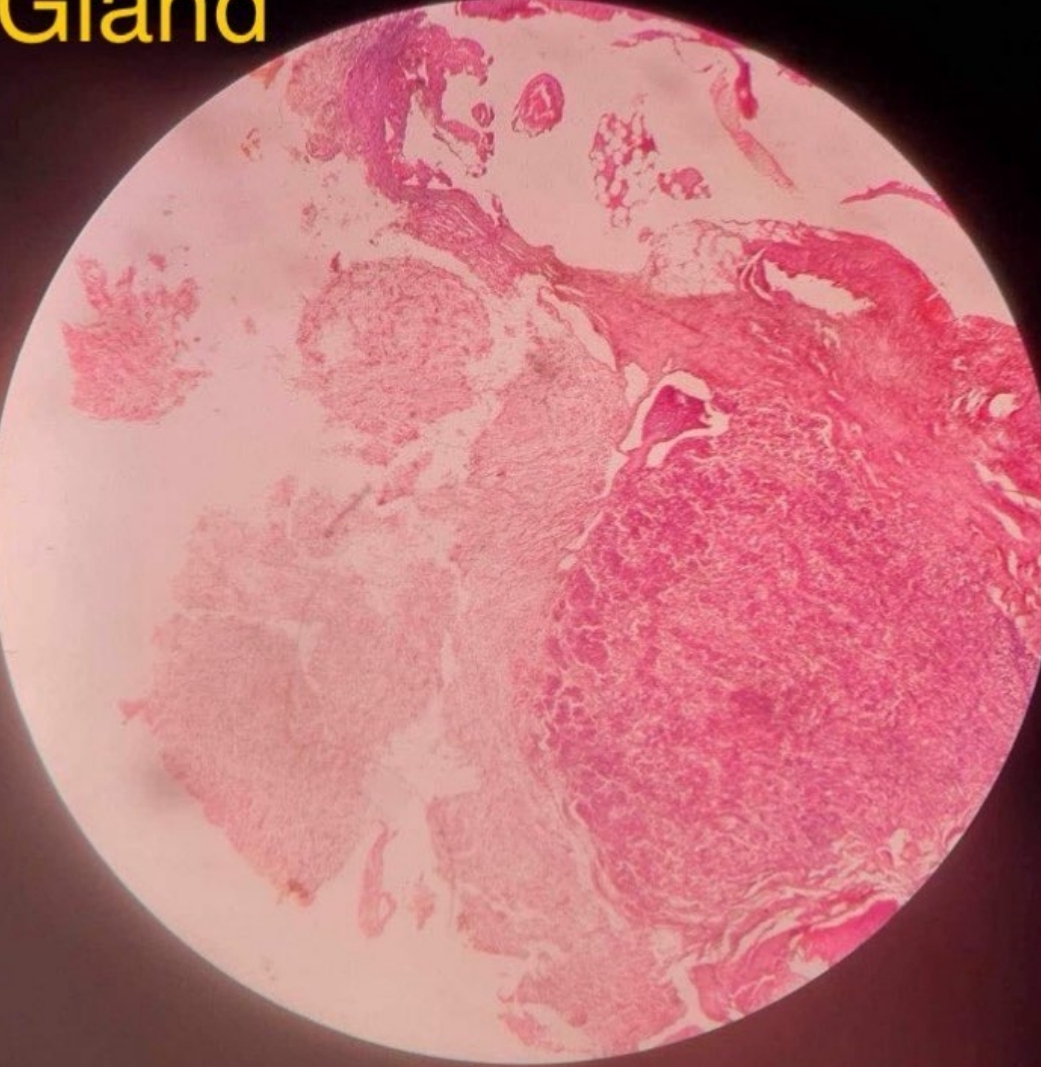
Parathyroid gland
Fig.
These glands are made up of masses of cells with numerous capillaries in between
Most of the cells (of which only nuclei are seen) are the chief cells which appear as small basophilic cells Oxyphil cells appear as large as eosinophilic (pink) cells
Oxyphil cells are few in number Adipose cells are also seen.
Adrenal gland
Fig.
The suprarenal gland is made up of a large number of cells arranged in layers.
It consists of an outer cortex and an inner medulla
The cortex is divisible into three zones
The zona glomerulosa is most superficial. Here, the cells are arranged in the form of inverted U-shaped structures or acinus-like groups In the zona fasciculata, the cells are arranged in straight columns (typically two-cell thick). Sinusoids intervene between the columns
The zona reticularis is made up of cords of cells that branch and form a network
The medulla is made up of groups of cells separated by wide sinusoids. Some sympathetic neurons are also present.
Reproductive system
Ovary
Fig.
The surface is covered by a cuboidal epithelium. Deep to the epithelium, there is a layer of connective tissue that constitutes the tunica albuginea
The substance of the ovary has an outer cortex in which follicles of various sizes are present, and an inner medulla consisting of connective tissue containing numerous blood vessels
Oviduct
Fig.
Fig.
The uterine tube is characterized by the presence of numerous branching mucosal folds that almost fill the lumen of the tube
The mucosa is lined by ciliated columnar epithelium
The uterine tube has a muscular wall with an inner circular and outer longitudinal muscle layer.
Uterus
Fig.
Fig.
Proliferative phase:
The wall of the uterus consists of a mucous membrane (called the endometrium) and a very thick layer of muscle (the myometrium). The thickness of the muscle layer helps to identify the uterus easily
The endometrium has a lining of columnar epithelium that rests on a stroma of connective tissue
Secretory phase:
The thickness of the endometrium is much increased
The uterine glands elongate, become dilated, and tortuous as a result of which they have saw-toothed margins in sections
Blood vessels extend in the upper portion of endometrium.
Vagina
Fig.
The vagina is a fibromuscular structure consisting of an inner mucosa, a middle muscular layer, and an outer adventitia
The mucosa consists of stratified squamous nonkeratinized epithelium and loose fibroelastic connective tissue lamina propria with many blood vessels and no glands
The mucosa of vagina is rich in glycogen and hence, the cells are pale stained which distinguishes it from esophagus
Muscular layer consists of smooth muscle fibers.
Testis
Fig.
Fig.
The testis has an outer fibrous layer, the tunica albuginea deep to which: A number of seminiferous tubules cut in various directions are seen
The tubules are separated by connective tissue, containing blood vessels and groups of interstitial cells of Leydig
Each seminiferous tubule is lined by several layers of cells
Cells are of two types:
Spermatogenic cells
Sustentacular
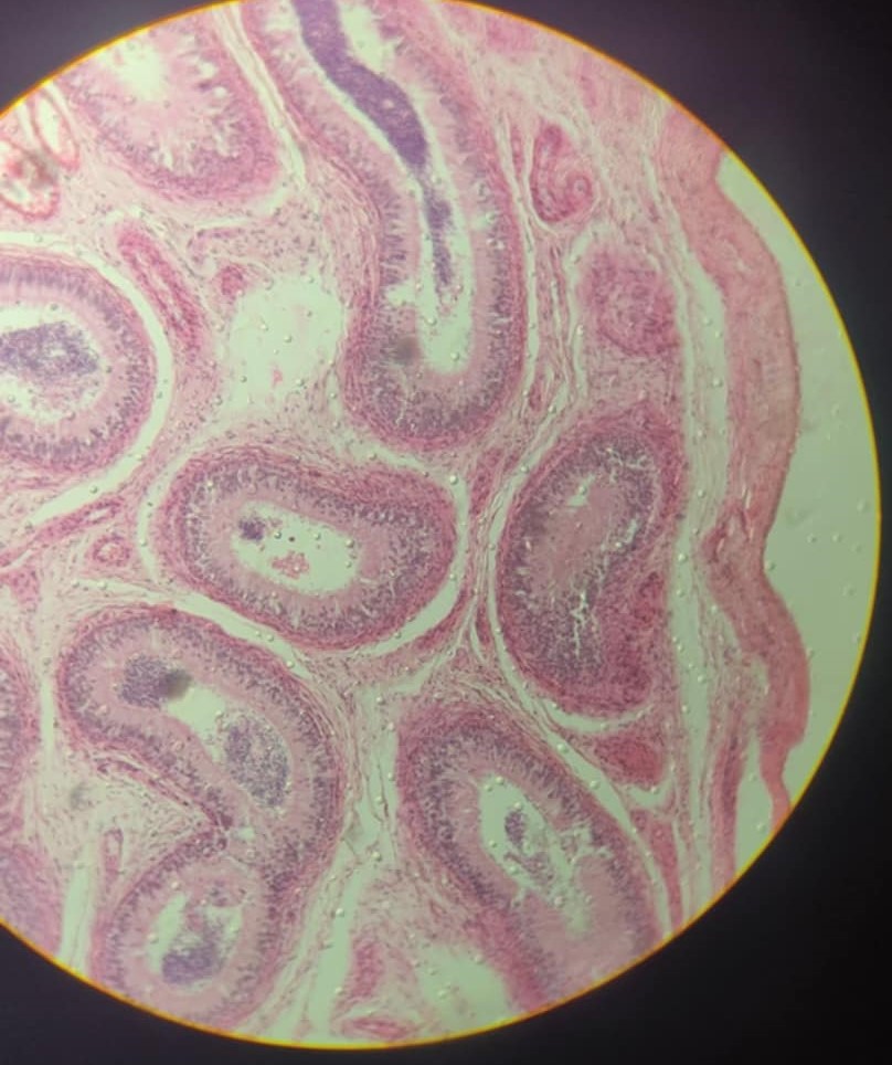
Epididymis
Fig.
The body of the epididymis is a long convoluted duct
A section shows number of tubules lined by pseudostratified columnar epithelium in which there are tall columnar cells and shorter basal cells that do not reach the lumen.
The columnar cells bear stereocilia
Smooth muscles are present in the wall of the duct
Clumps of spermatozoa are present in the lumen of the duct.
Vas deferens
Fig.
Fig.
This tubular structure displays: A small irregular lumen
Mucous membrane lined by pseudostratified columnar epithelium with underlying lamina propria
The muscle coat is very thick. Three layers, inner longitudinal, middle circular, and outer longitudinal are seen
Outer most layer is adventitia composed of collagen fibers and containing blood vessels.
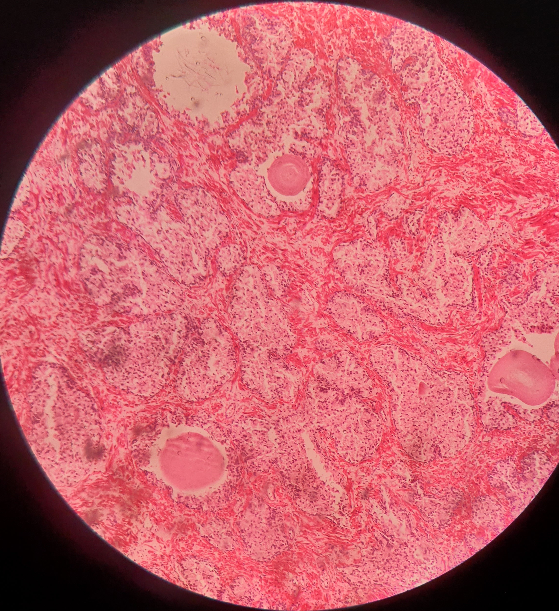
Prostate
Fig.
Fig.
The prostate consists of glandular tissue embedded in prominent fibromuscular stroma
The glandular tissue is in the form of follicles with serrated edges. They are lined by columnar epithelium. The lumen may contain amyloid bodies
The follicles are separated by broad bands of fibromuscular tissue.
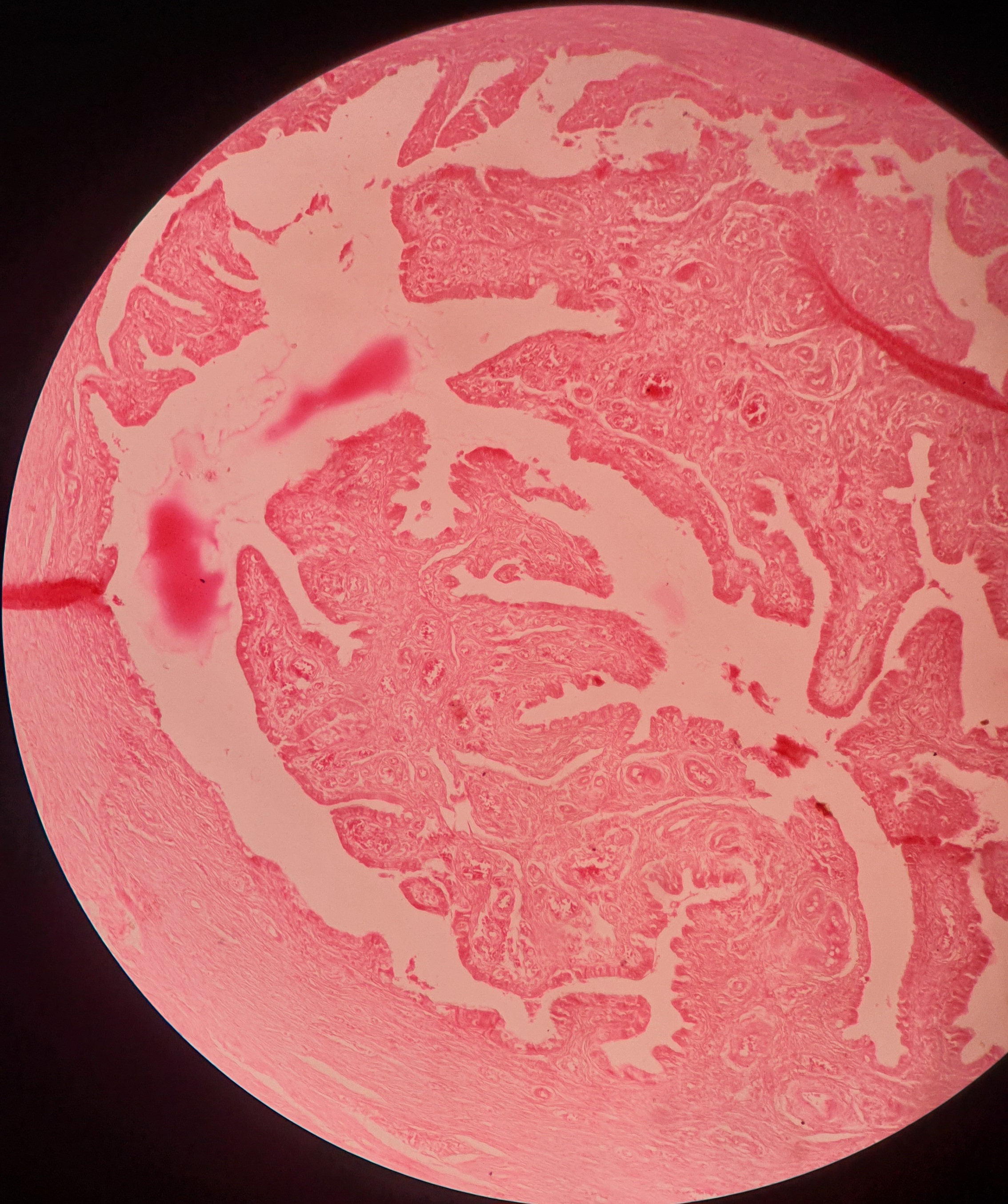
Seminal vesicle
Fig.
The seminal vesicle is made up of a convoluted tubule
The tube has an outer covering of connective tissue, a thin layer of smooth muscle and an inner mucosa
The mucosal lining is thrown into numerous folds that branch and anastomose to form a network
The lining epithelium is usually simple columnar or pseudostratified.
Penis
Fig.
Fig.
Fig.
The testis has an outer fibrous layer, the tunica albuginea deep to which:
A number of seminiferous tubules cut in various directions are seen
The tubules are separated by connective tissue, containing blood vessels and groups of interstitial cells of Leydig
Each seminiferous tubule is lined by several layers of cells
Cells are of two types:
Sustentacular cells
Spermatogenic cell
Renal system
Kidney
Fig.
The kidney is covered by a capsule
Deep to the capsule there is the cortex
Deep to the cortex there is the medulla of the kidney
In the cortex we see circular structures called renal corpuscles surrounding which there are tubules cut in various shapes
The dark pink stained tubules are parts of the proximal convoluted tubules (PCT): their lumen is small and indistinct. It is lined by cuboidal epithelium with brush border
Lighter staining tubules, each with a distinct lumen, are the distal convoluted tubules (DCT).
They are lined by simple cuboidal epithelium PCT are more in number than DCT
In the medulla we see very light staining, elongated, parallel running tubules.
These are collecting ducts and loop of Henle. Some of them extend into the cortex forming a medullary ray. The collecting ducts are lined by simple cuboidal epithelium and loop of Henle (thin segments) are lined by simple squamous epithelium
Cut sections of blood vessels are seen both in the cortex and medulla.
Ureter
Fig.
The ureter can be recognized because it is tubular and its mucous membrane is lined by transitional epithelium
The epithelium rests on a layer of connective tissue (lamina propria)
The mucosa shows folds that give the lumen a star-shaped appearance
The muscle coat has an inner layer of longitudinal fibers and an outer layer of circular fibers.
This arrangement is the reverse of that in the gut
The muscle coat is surrounded by connective tissue–adventitia in which blood vessels and fat cells are present.
Urinary bladder
Fig.
Fig.
The urinary bladder is easily recognized because the mucous membrane is lined by transitional epithelium
The epithelium rests on lamina propria
The muscle layer is thick. It has inner and outer longitudinal layers between which there is a layer of circular or oblique fibers.
The distinct muscle layers may not be distinguishable
The outer surface is lined in parts by peritoneum (serosa)