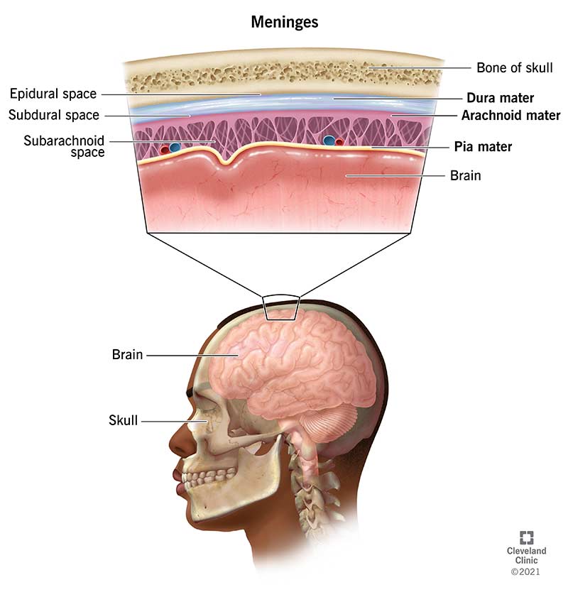
1. General Anatomy
  2. 1. Introduction
    2. Skeleton
    3. Joints
    4. Muscles
    5. Cardiovascular system
    6. Lymphatic system
    7. Nervous system
    8. Skin and Fasciae
    9. Connective tissue, Ligaments and Raphe
    10. Principles of Radiography
  3. Head and Neck
  4. 11. Introduction and Osteology
    12. Scalp, Temple and Face
    13. Anterior Triangle of the Neck
    14. Parotid Region
    15. Temporal and Infratemporal Regions
    16. Submandibular region
    17. Structures in the Neck
    18. Prevertebral and Paravertebral Regions
    19. Back of the Neck
    20. Contents of the Vertebral Canal
    21. Cranial Cavity
    22. Contents of the Orbit
    23. Mouth and Pharynx
    24. Nose, Paranasal Sinuses and Pterygopalatine Fossa
    25. Larynx
    26. Tongue
    27. Ear
    28. Eyeball
    29. Surfacemarking and Radiological Anatomy
  5. Neuroanatomy
  6. 30. Introduction
    31. Meninges of the Brain and Cerebrospinal Fluid
    32. Spinal cord
    33. Cranial nerves
    34. Brainstem
    35. Cerebellum
    36. Fourth Ventricle
    37. Cerebrum, Diencephalon, Basal nuclei and White matter
    38. Third ventricle, Lateral ventricle and Limbic system
    39. Some neural pathways nad Reticular formation
    40. Blood supply of Spinal cord and Brain
    41. Investigations of a Neurological Case, Surface marking
    42. Autonomic Nervous System
  7. Upper limb
  8. 43. Introduction
    44. Bones
    45. Pectoral region
    46. Axilla
    47. Back
    48. Scapular region
    49. Cutaneous nerves, Superficial veins and Lymphatic drainage
    50. Arm
    51. Forearm and Hand
    52. Joints of Upper limb
    53. Surface marking, Radiological anatomy
  9. Thorax
  10. 54. Introduction
    55. Bonesa and Joints of Thorax
    56. Walls of Thorax
    57. Thoracic cavity and Pleurae
    58. Lungs
    59. Mediastinum
    60. Pericardium and Heart
    61. Superior venacava, Aorta and Pulmonary trunk
    62. Trachea, Oesophagus and Thoracic duct
    63. Surface marking and Radiological anatomy of Thorax
  11. Abdomen and Pelvis
  12. 64. Introduction and Osteology
    65. Anterior Abdominal Wall
    66. Male External Genital Organs
    67. Abdominal cavity and Peritoneum
    68. Abdominal part of Oesophagus and Stomach
    69. Small and Large intestines
    70. Large Blood vessels of the Gut
    71. Extrahepatic Biliary Apparatus
    72. Spleen, Pancreas and Liver
    73. Kidney and Ureter
    74. Suprarenal gland and Chromaffin system
    75. Diaphragm
    76. Posterior abdominal wall
    77. Perineum
    78. Preliminary consideration of Boundaries and Contents of Pelvis
    79. Urinary Bladder and Urethra
    80. Female Reproductive Organs
    81. Male Internal Genital Organs
    82. Rectum and Anal Canal
    83. Walls of Pelvis
    84. Surface marking of Abdomen and Pelvis
    85. Radiological and Imaging procedures
  13. Lower limb
  14. 86. Introduction
    87. Bones
    88. Front of Thigh
    89. Medial side of Thigh
    90. Gluteal region
    91. Popliteal fossa
    92. Back of Thigh
    93. Front of Leg with Dorsum of Foot, Lateral and Medial sides of Leg
    94. Back of Leg
    95. Sole of Foot
    96. Venous and Lymphatic drainage; Segmental adn Sympathetic Innervations; and Comparison of Lower and Upper Limbs
    97. Joints of Lower Limb
    98. Arches of Foot
    99. Surface and Radiological anatomy
Neuroanatomy
31. Meninges of the Brain and Cerebrospinal Fluid
Image 1
Credit. Clevland cleinic
THE MENINGES
Duramater
Arachnoid mater
Piamater
Character
Duramater
Arachnoid mater
Pia mater
Etmology
Dura -> Hard mother
Arachnoid -> Cobweb like
Pia -> Soft mother
Layers
Endosteal layer:
- Cranial venous sinuses are present between these two layers
Meningeal layer:
Folds:
Character
Falx cerebri
Falx cerebelli
Tentorium cerebelli
Diaphragma sellae
Shape
Sickle-shaped
Small sickle-shaped
Tent-shaped
Small horizontal fold
Functions
Separates the right from left cerebral hemisphere
Partly separates the cerebellar hemispheres
  1. Separates the cerebral hemispheres from hindbrain and lower part of midbrain
  2. Lifts off the weight of occipital lobes from the cerebellum.
Venous sinuses enclosed
  1. Superior sagittal sinus
  2. Inferior sagittal sinus
  3. Straight sinus
Occipital sinus
  1. Transverse sinuses
  2. Superior petrosal sinuses
Anterior and posterior intercavernous sinuses
Location
Just beneath the skull
Below the Duramater
Below the Arachnoid mater, covering the brain
Spaces
Epidural space: Space just above the duramater (Between Skull and Duramater)
Subdural space: Space below the duramater (Between duramater and arachnoid mater)
Subarachnoid space:
  • Space just below the arachnoid mater. (Between Arachnoid and Pia mater)
  • CSF (Cerebrospinal fluid) flows through Subarachnoid space.
No spaces