Stains poorly with H&E, special stains like orcein
Black with silver stain (argyrophilic)
4
Function
Tensile strength (resist stretching)
Elasticity (return to shape after stretch)
Form supportive mesh in soft tissues
5
Location
Type I
Type II
Type III
Type IV
Type VI
Bones
Tendons
Ligaments
Skin
Hyaline cartilage
Liver
Bone marrow
Lymphoid organs
Basement membrane
Basement membrane
Lungs, arteries, skin, elastic cartilage
Lymph nodes, spleen, bone marrow
❖ Matrix/ Ground substance:
Connective tissue proper
Loose connective tissue
Areolar connective tissue
Adipose connective tissue
Dense connective tissue
Fibrous connective tissue
White fibrous connective tissue
Tendon
Ligaments
Yellow elastic connective tissue
Reticular connective tissue
Pigmented connective tissue
Fluid connective tissue
Blood
▢ Blood plasma:
▢ Blood cells:
❖ Red Blood Cells (RBCs)/ Erythrocytes:
❖ White Blood Cells (WBCs)/ Leucocytes:
Granulocytes
Agranulocytes
With Cytoplasmic granules.
They have lobed nucleus.
Without cytoplasmic granules.
They don't have lobed nucleus.
Eosinophils
Basophils
Neutrophils
Monocytes
Lymphocytes
Plasma cell
2 nucleus-lobe
2-3 nucleus-lobe
3-5 nucleus-lobe
Largest WBC
Smallest WBC
Phagocytic
Non-phagocytic
Phagocytic
Phagocytic
Involved in Body defence mechanism
Involved in Allergic reaction
Secrete histamine and Serotonin
First line of defense against infection.
Form Macrophages in tissues.
◉ Granulocytes:
◈ Eosinophils:
◈ Basophils:
◈ Neutrophils:
◉ Agranulocytes:
◈ Monocytes:
■ Macrophages in tissues:
Brain → Microglia
Skin → Dendritic cells/ Langerhans' cells
Connective tissue → Histiocytes
Cartilage → Condrocytes
Bone → Osteocytes
Thymus → Hassal's corpuscles
Lungs → Dust cells
Liver → Kuffer cells
Spleen → Sinusoidal cells
Kidney → Messangial cells
◈ Lymphocytes:
■ T-lymphocytes:
■ B-lymphocytes:
◈ Plasma cells:
❖ Platelets/ Thrombocytes:
Lymph
Skeletal connective tissue
Bone
Cartilage
MUSCULAR TISSUE
Skeletal muscle
Smooth muscle
Cardiac muscle
NERVOUS TISSUE
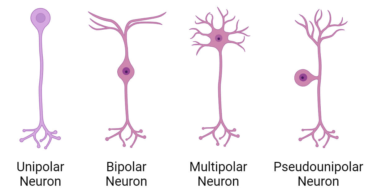
Neurons
Apolar neuron
Unipolar neuron
Pseudo-unipolar neuron
Bipolar neuron
Multipolar neuron
Location
Hydra
Some Amacrine cells of Retina
Chromaffin cells
Vertebrate embryo
Trigeminal nerve nucleus
a. Dorsal root ganglia of Spinal cord i.e, most sensory nerve
Retina (Eye)
Cochlea (Ear)
Olfactory area (Nose)
General (Brain)
Autonomic ganglia
Purkinje cells
Fea
Glial cells
Astrocytes
Oligodendrocytes
Microglia
Ependymal cells
Satellite cells
Schwan cells
Location
CNS
PNS
Functions
Astrocytes
Oligodendrocytes
Microglia
Ependymal cells
Satellite cells
Schwan cells
1. Active secretary cells in human body are
[MOE 2069]
Myoblast
Goblet cells
Monocytes
Erythrocytes
(b) Goblet cells are active secretory cells, particularly in mucous membranes.
2. Histamine is secreted by:
[BPKIHS 2012]
Lymphocyte
Mast cell
Osteocyte
RBC
(b) Histamine is primarily secreted by mast cells.
3. Keratinised stratified squamous epithelium is found in
[IOM 2010]
Trachea
Lining of blood vessel
Mouth cavity
Epidermis
(d) Keratinized stratified squamous epithelium is found in the epidermis (skin).
4. The acidophils are
[MOE 2010]
White blood cells
Connective tissue cells
Bone cells
Cartilage cells
(a) Acidophils (now called eosinophils) are a type of white blood cell.
5. Dendron or dendrite is a part of
[MOE 2010]
Connective tissue
Nervous tissue
Epithelial tissue
Skeletal tissue
(b) Dendrites are part of a neuron (nervous tissue).
6. Which one of the human cell do not contain mitochondria
[KU 2012]
Nerve cell
Red blood cells
Liver cells
White blood cells
(b) Mature red blood cells lack mitochondria.
7. Urinogenital ducts are lined by
[KU 2010]
Pseudostratified columnar epithelium
Glandular epithelium
Simple squamous epithelium
Stratified squamous epithelium
(a) Urinogenital ducts are lined by Pseudostratified columnar epithelium.
8. The tissue that protect body from wound and infection is
[MOE 2011, 2008/KU]
Areolar
Adipose
Reticular tissue
Lymphatic
(d) Lymphatic tissue is part of the immune system, protecting against infection.
9. Serum is
[IOM 2010]
Plasma
Plasma minus Calcium ions
Plasma minus Fibrinogen
Plasma minus Gamma globulins
(c) Serum is plasma without fibrinogen.
10. Serum is:
[BPKIHS 2012]
Blood-plasma
Plasma-blood cells
Plasma-cIotting factors
Plasma-WBC
(c) Serum is plasma without clotting factors.
11. The skeleton and muscles are derived from:
[MOE 2008/IOM 2010]
Ectoderm and mesoderm
Ectoderm
Endoderm
Mesoderm
(d) The skeleton and muscles are derived from the mesoderm.
12. The bone of mammals contains longitudinal Haversian canals, which are connected by transverse canals called
[IOM 2010]
Bidder's canal
Volkmann's canal
Inguinal canal
Eustachian canal
(b) Volkmann's canals connect Haversian canals in bone.
13. Which of the following leukocytes doesn't contain granules in cytoplasm?
[KU 2009]
Neutrophil
Basophil
Monocyte
Eosinophil
(c) Monocytes are agranulocytes (lack cytoplasmic granules).
14. Mast cell in the connective tissue resemble
[MOE 2009]
Basophils
Monocytes
Plasma cells
Sertoli cells
(a) Mast cells are similar to basophils in function and appearance.
15. The cells which do not divide:
[MOE 2060, 2008]
Neuron
Muscular
Blood cell
Bone cell
(a) Neurons (nerve cells) generally do not divide.
16. Histamine and Heparin are secreted by:
[IE 2008]
Plasma cell
Mast cell
Lymphocyte
Macrophages
(b) Mast cells secrete both histamine and heparin.
17. Nissil's granules are found in:
[IE 2008]
Axon
Myofibrill
Nerve Cell
Mylenated sheath
(c) Nissl's granules are found in the cell body of nerve cells.
18. Transitional epithelium is found in
[IE 2008, IOM 1993]
Gall bladder
Liver
Urinary bladder
Vagina
(c) Transitional epithelium is located in the urinary bladder.
19. The internal lining of the blood vessel is:
[MOE 2064]
Mesothelium
Endothelium
Pavement epithelium
Stratified epithelium
(b) The inner lining of blood vessels is endothelium.
20. The fibrous sheath that connects the bone is called:
[MOE 2064, 2061]
Tendon
Aponeurosis
Periosteum
Ligament
(d) Ligaments connect bone to bone.
21. Outer covering of bone is:
[MOE 2063]
Perichondrium
Endosteum
Periosteum
Pericardium
(c) The outer covering of bone is the periosteum.
22. What is the name of diseases caused by the endocrinology deficiency of thyroxine
[MOE 2062]
Diabetes
Night blindness
Goitre
Addison's diseases
(c) Deficiency of thyroxine leads to Goitre.
23. 'Reticulin fibres' are associated with
[MOE 2063]
Retina
Reticulocytes
Phagocytosis
All of the above
(c) Reticulin fibers are involved in supporting cells and phagocytosis.
24. What is the function of motor nerves?
[MOE 2062]
Conduction of impulse to CNS
Conduction of impulse away from CNS
Both
None
(b) Motor nerves conduct impulses away from the CNS.
25. Yellow bone marrow is found especially in the medullary cavity of
[MOE-2062]
Spongy bones
Long bones
Short bones
All of these
(b) Yellow bone marrow is found in the medullary cavity of long bones.
26. Memory cells are
[MOE-2061]
T cells only
B cells only
T and B cells both
None
(c) Both T and B cells can become memory cells.
27. The growth of hair follicle is facilitated by
[MOE-2060]
Stratum corneum
Stratum malpighii
Dermis
None
(c) The dermis supports hair follicle growth.
28. Which of the following is anticoagulant
[IOM 2005]
Heparin
Protein
Fibrinogen
Thromboplastin
(a) Heparin is an anticoagulant.
29. A very good power of regeneration is found in
[BP 2007]
Lung
Liver
Kidney
Spleen
(b) The liver has a high capacity for regeneration.
30. Blood vessels near a wound dilate & become more permeable in response to which materials release from damaged cells?
[IE 2007]
Pyrogens
Antibodies
Histamine
Interferon
(c) Histamine released from damaged cells increases blood vessel permeability.
31. Abdominal organs are covered by
[BP 2006]
Peritoneum
Omentum
Pericardium
Pleura
(a) The peritoneum covers abdominal organs.
32. Which of the following increases during allergy?
[BPKIHS 2006]
IgE
IgA
IgM
IgD
(a) IgE levels increase during allergic reactions.
33. Trachea is lined by
[BPKIHS 2006]
Pseudo stratified columnar epithelium
Stratified epithelium
Cuboidal epithelium
Squamous epithelium
(a) The trachea is lined by pseudo stratified columnar epithelium.
34. Intercalated discs are found in
[BPKIHS 2005]
Cardiac muscle
Smooth muscles
Striated muscles
Ligaments
(a) Intercalated discs are characteristic of cardiac muscle.
35. Inflammation characterized by dilation of capillaries & small blood vessels surrounding the injuries infected area becomes red, warm & swollen due to increase permeability to
[IE 2005]
Heparin
Histamine
Macrophage
Interferon
(b) Histamine increases capillary permeability during inflammation.
36. Heparin is formed by
[IE 2004]
Mast cell
Kidney cells
Blood cells
Bone marrow cells
(a) Heparin is produced by mast cells.
37. WBC that are non-specific killer of microbes are
[IE - 2004]
B-cell
Phagocyto
Killer T
Helper T
(b) Phagocytes are non-specific killers of microbes.
38. Wrinklenge of skin in old age is due to
[BP 2004)]
Collagen
Keratin
Action
Myosin
(a) Wrinkling in old age is due to loss of collagen.
39. Cells of germinal epithelium are
[BPKIHS 2007]
Columnar
Glandular
Cuboidal
Ciliated
(c) Germinal epithelium cells are cuboidal.
40. A very good power of regeneration is found in
[BPKIHS 2007]
Lung
Liver
Kidney
Spleen
(b) The liver has a high capacity for regeneration.
41. Tendon connects:
[MOE 2002)]
Bone to bone
Bone to muscle
Muscle to muscle
Bones of cranium
(b) Tendons connect bone to muscle.
42. Protoplasmic connection between two cells is
[MOE 2002]
Plasmodesmata
Cell wall
Plasma membrane
Cell membrane
(a) Plasmodesmata are protoplasmic connections between plant cells. Gap junctions are the equivalent in animal cells, but the term isn't in the options.
43. Which of the following may arise from any 3 germ layers? [[BPKIHS 2001]
[BPKIHS 2001]
Epithelial tissue
Muscular tissue
Connective tissue
Nervous tissue
(a) Epithelial tissue can arise from all three germ layers (ectoderm, mesoderm, and endoderm).
44. A group of cell having common origin and similar function is [MOE 2000]
[MOE 2000]
Tissue
System
Organ
Body
(a) A tissue is a group of cells with a common origin and similar function.
45. Cardiac muscles are found in
[MOE 2056]
Kidney of frog
Liver of frog
Heart of mammal
Stomach of rabbit
(c) Cardiac muscle is found in the heart of mammals.
46. The substance produced in the body for defence is
[BPKIHS 1998]
Platelets
Antigens
Lymphocytes
Thrombins
(c) Lymphocytes are white blood cells involved in the immune response (defense).
47. Fat cells are [IOM 1997]
[IOM 1997]
Epithelial tissue
Connective tissue
Muscular tissue
Adipose tissue
(d) Fat cells are a type of adipose tissue, which is a connective tissue.
48. Which of the following is the retroperitoneal organ?
[BP 2000]
Spleen
Testes
Heart
Kidney
(d) The kidneys are retroperitoneal, located behind the peritoneum.
49. Irreversible cell death is known as:
[BPKIHS 1995]
Necrosis
Degeneration
Anaplasia
Fibrosis
(a) Necrosis is irreversible cell death.
50. Glisson's capsule is found in which mammalian organ?
[BP 1994]
Kidney
Liver
Pancreas
Brain
(b) Glisson's capsule is the connective tissue capsule surrounding the liver.
51. Glands and ducts are internally lined with
[IOM 1993]
Columnar epithelium
Cuboidal epithelium
Ciliated columnar epithelium
Transitional epithelium
(b) Glands and ducts are typically lined with cuboidal epithelium.
52. Ciliated epithelium is NOT found in
[IOM 1993]
Respiratory tract
Fallopian tube
Vas deferens
Ureter
(c) Ciliated epithelium is not found in the vas deferens.
53. In which of the following, ciliated epithelium is present?
[BPKIHS -2013]
Trachea
Oesophagus
Ileum
Stomach
(a) Ciliated epithelium is present in the trachea.
54. Fat is abundant in :
[BPKIHS -2013]
Areolar tissue
Adipose tissue
Connective tissue
Muscular tissue
(b) Fat is abundant in adipose tissue.
55. in diapedesis, which of the following squeezes out of the narrow capillaries? [IOM-2014]
[IOM-2014]
RBC
Plasma
Platelets
WBC
(d) White blood cells (WBCs) undergo diapedesis.
56. The ridges and finger prints in the hands is due to [IOM-2014]
[IOM-2014]
Dermal papillae
Stratum lucidium
Stratum granulosum
Stratum germinativum
(a) Fingerprints are due to dermal papillae.
57. What is insoluble in water?
[BPKIHS -2013]
α-keratin
Myoglobin
Globulin
Iodochromatin
(a) α-keratin is insoluble in water.
58. Clotting of blood is delayed by:
[IOM-2013]
Prothrombin
Heparin
Vitamin K
Calcium
(b) Heparin is an anticoagulant that delays blood clotting.
59. Connective tissue is originated from :
[IOM-2013]
Endoderm
Mesoderm
Ectoderm
Ecto-mesoderm
(b) Connective tissue originates from the mesoderm.
60. Histamine is secreted by:
[IOM-2013]
Mast cell
Osteoblast
Fibroblast
Macrophage
(a) Histamine is secreted by mast cells.
61. The membrane that covers the cartilage is known as:
[IOM-2013]
Perichondrium
Periosteum
Pericardium
Perineurium
(a) The perichondrium covers cartilage.
62. Transitional epithelium is found in:
[IOM-2013)]
Urinary bladder
Liver
Kidney
Oesophagus
(a) Transitional epithelium is found in the urinary bladder.
63. Blood is:
[IOM-2013]
Connective tissue
Muscular tissue
Epithelial tissue
Nervous tissue
(a) Blood is a type of connective tissue.
64. Cartilage present between the joint of long bone is:
[BPKIHS -2014,015]
Fibrous cartilage
Hyaline
Elastic
None of above
(b) Hyaline cartilage is found in the joints of long bones.
65. Which mineral is needed for muscle contraction:
[BPKIHS -2014]
Ca
Mg
Lactic Acid
All
(a) Calcium (Ca) is essential for muscle contraction.
66. Muscle become fatigue due to excess accumulation of: